
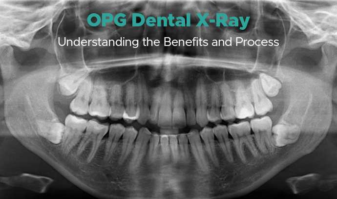
OPG Dental X-Ray: Understanding the Benefits and Process
Patients are often referred for an OPG scan without a detailed explanation of its purpose. An OPG dental x-ray, also called an orthopantomogram or panoramic x-ray, is a comprehensive radiography procedure that captures the teeth, jaws, roots, and surrounding oral structures in a single image. It is routinely recommended for the evaluation, screening, and treatment planning of orthodontic, implant, and surgical cases.
At Elite Dental Studio, Calicut, advanced digital radiology equipment and experienced specialists ensure accurate, safe, and comfortable OPG dental x-ray. Read this blog till the end to know all about the benefits, process, and safety measures involved in an OPG examination.
What Is an OPG Dental X-Ray?
An OPG (Orthopantomogram) is a panoramic imaging procedure that records a two-dimensional view of the oral and maxillofacial region. During the scan, the x-ray machine rotates gently around the patient’s head while a digital sensor or detector captures continuous images, later combined into a single panoramic view.
The resulting image displays the teeth, gums, jawbone, sinus, roots, nerves, and temporomandibular joints. This wide coverage allows dentists to assess tooth alignment, arch structure, bite, and overall oral health. OPG imaging is widely used in orthodontics, implantology, periodontics, endodontics, and maxillofacial surgery for precise diagnosis and treatment planning.
Benefits of OPG
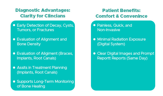
An OPG scan offers both diagnostic and patient-centric advantages, making it an essential part of comprehensive dental care.
Diagnostic Advantages
An OPG provides complete visibility across both jaws, enabling early detection of decay, infection, cysts, tumours, fractures, or lesions. It assists clinicians in evaluating alignment, bone density, and pathological changes with clarity and accuracy.
Treatment Planning and Monitoring
Dentists use OPG images to design treatment plans for braces, implants, root canal therapy, and extractions. The scan supports long-term monitoring of bone healing, jaw stability, and changes after orthodontic or surgical care.
Patient Benefits
The scan is painless, quick, and non-invasive. It requires minimal radiation exposure, produces clear digital images, and delivers reports promptly. These features make it convenient for routine screening and advanced diagnostic evaluation.
The OPG Process, Step by Step
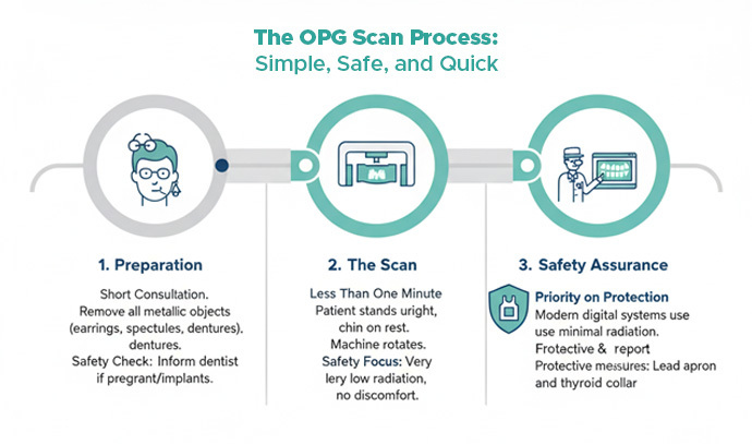
Understanding how the scan works helps patients know what to expect during the appointment.
1. Pre-Examination Preparation
Before the procedure, the dental team conducts a short consultation and assessment. Patients are asked to remove any metal objects, such as earrings, spectacles, or removable dentures, to avoid image distortion. This simple preparation step ensures accurate capture and high image clarity.
2. During the Scan
The patient stands upright with the chin placed on a rest and gently bites on a sterile block. The machine completes a controlled rotation around the head while the sensor records the exposure. The process takes less than one minute and causes no discomfort.
3. Post-Examination Review
After the scan, the digital image is displayed immediately on the system. A radiologist or dentist performs an analysis, prepares the report, and explains the findings during the consultation. The image serves as a valuable reference for further diagnostic evaluation or treatment planning.
OPG Dental X-Ray: Safety and Radiation
Patient safety is a priority during all radiographic procedures at Elite Dental Studio, and OPG imaging follows strict protection protocols.
Controlled Exposure
Modern digital radiography systems use very low radiation doses, significantly less than traditional film-based methods. Advanced technology ensures sharp image resolution while maintaining patient protection.
Protective Measures
Standard safety equipment such as a lead apron and thyroid collar is provided when necessary. Proper positioning and machine calibration reduce the need for repeat scans, maintaining minimal overall exposure.
Important Precautions
Patients who are pregnant, have undergone recent imaging, or have medical implants should inform the dentist in advance. These details allow the clinician to adjust the procedure or postpone it when required.
OPG Compared with Other Dental Imaging Methods
Different imaging methods serve specific purposes. The OPG stands out for its ability to capture an overall view of the oral cavity efficiently.
OPG vs Intraoral Radiography
Intraoral x-rays focus on small areas, capturing one or two teeth for detailed examination. They help detect fine cracks or caries. An OPG, in contrast, provides a complete panoramic overview of both jaws, offering insight into structural relationships and tooth alignment.
OPG vs CBCT
A CBCT (Cone Beam Computed Tomography) scan delivers a three-dimensional view useful for implant placement, orthodontic assessment, or maxillofacial surgery. An OPG is often used first for general screening, while CBCT is chosen for cases needing 3D analysis and volumetric detail.
Integrated Diagnostic Approach
Clinicians may combine OPG, CBCT, and intraoral imaging to achieve complete evaluation and accurate treatment planning. This balanced approach offers both detail and context while limiting radiation exposure.
OPG Dental X-Ray: Results and Interpretation
After the scan, the radiologist examines the image to identify tooth structure, root formation, bone levels, nerve pathways, and potential pathology such as infection, fracture, or anomaly. A concise report is then shared with the treating dentist.
This information supports accurate decisions for orthodontic adjustments, implant placement, root canal therapy, or tooth extraction. Precise interpretation ensures effective planning and improved clinical outcomes.
OPG Dental X-Ray: Patient Guidelines for an Effective Examination
Following a few practical measures helps achieve optimal image quality and a smooth experience.
- Arrive a few minutes before the appointment to complete registration.
- Remove metallic items and accessories to avoid image artefacts.
- Follow positioning instructions carefully and remain still during the rotation.
- Maintain normal breathing to prevent motion blur.
- Request a copy of the digital image or report for your records.
These steps ensure clear imaging, efficiency, and consistent diagnostic accuracy.
Book Your OPG Dental X-Ray with Elite Dental Studio, Calicut Now
Elite Dental Studio provides advanced digital panoramic imaging with a focus on accuracy, safety, and patient comfort. Each OPG scan is supervised by qualified radiology specialists and reviewed by senior dentists for reliable results.
Appointments are easily available, with same-day reports and prompt consultations. To schedule your OPG Dental X-Ray, contact your nearest Elite Dental Studio, Calicut
Whether you’re searching for a dental clinic in Calicut or a dental hospital in Kochi, Elite Dental Studio in Calicut offers a range of dental treatments under one roof. Book a Consultation with our experts now.
Frequently Asked Questions (FAQ)
Is an OPG dental x-ray painful?
No. The OPG dental x-ray is a non-invasive and painless radiography procedure. The panoramic scan completes in less than a minute with no discomfort.
How long does the OPG process take?
The full OPG dental x-ray process takes about five minutes, including positioning and capture. The actual imaging lasts under sixty seconds.
Is radiation exposure high in an OPG scan?
No. Modern digital radiography equipment uses minimal radiation exposure. Standard protection protocols, such as a lead apron, ensure complete safety.
Is an OPG required before braces or dental implants?
Yes. An OPG scan helps evaluate jaw structure, bone density, root alignment, and tooth positioning, which are essential for braces or implant planning.
Can I receive my OPG report on the same day?
Yes. The digital OPG image appears instantly, and the radiologist provides the diagnostic report and interpretation within a few hours of the scan.
What conditions can an OPG detect?
An OPG dental x-ray identifies decay, infection, cysts, tumours, fractures, and anomalies in the teeth, jawbone, and oral structures accurately.
Can children undergo an OPG scan safely?
Yes. When clinically required, pediatric OPG imaging is performed using adjusted exposure settings and radiation protection to ensure complete safety.
Have Dental Problem : Call us
CALICUT: +91 9745 072 555,
KOCHI: +91 9567 124 888
KANNUR: +91 9645874777
or make an Appointment
Take a smiling selfie and we'll Simulate your new smile See What Invisalign treatment could do for you!